
Figure 1.
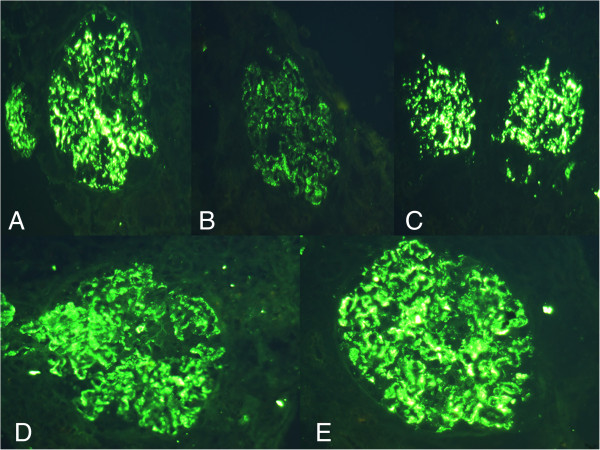
Immunofluorescence microscopy showed strong staining of (A) IgG, (B) IgA, (C) C3, (D) IgG kappa, and (E) IgG lambda over mesangium and glomerular basement membrane (original magnification, × 400).
Official websites use .gov
A
.gov website belongs to an official
government organization in the United States.
Secure .gov websites use HTTPS
A lock (
) or https:// means you've safely
connected to the .gov website. Share sensitive
information only on official, secure websites.
Immunofluorescence microscopy showed strong staining of (A) IgG, (B) IgA, (C) C3, (D) IgG kappa, and (E) IgG lambda over mesangium and glomerular basement membrane (original magnification, × 400).