
Figure 1.
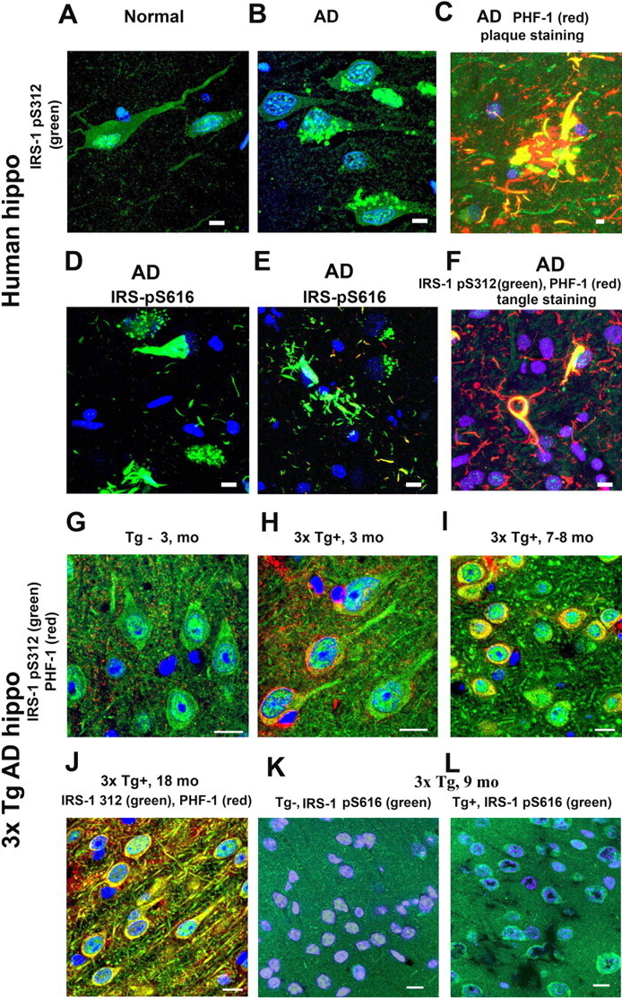
Redistribution of phosphorylated IRS-1 in AD and in 3xTg-AD mice and its enhancement by high-fat bad diet in 3xTg-AD mice. Using an antibody to the pS312 phosphoepitope of IRS-1, confocal immunocytochemistry illustrated that neurons from control human hippocampus (hippo) expressed high levels in the nucleus (A), whereas in AD patients, it was redistributed in the cytosol, sometimes in a granulovacuolar pattern (B). Around plaques, increased IRS-1 staining colocalized with dystrophic neurites (C). Antibodies to phosphorylated IRS-1 Ser616 typically stained tangles (D) and dystrophic neurites (E). IRS-1 pS312 colocalized with PHF-1 (F). Compared with 3-month-old control mice, which showed hippocampal CA1 nuclear staining of IRS-1 pS312 (G), 3xTg-AD mice had elevated IRS-1 pS312 in cytosol (H), which increased at 7–8 months of age (I). By 18 months of age, there was extensive colocalization of IRS-1 ps312 with PHF-1 (J). This transgene effect was also seen in animals using another IRS-1 antibody (pS616) in an experiment in which 5-month-old 3xTg-AD mice were fed with a HFBD (see text) for 4 months, and the phosphorylation of IRS-1 Ser616 was examined. Compared with Tg− control mice (K), 3xAD-Tg+ mice showed more cytosolic pS616 IRS-1 labeling and minimal nuclear labeling at 9 months of age (L). Scale bars, 20 μm.