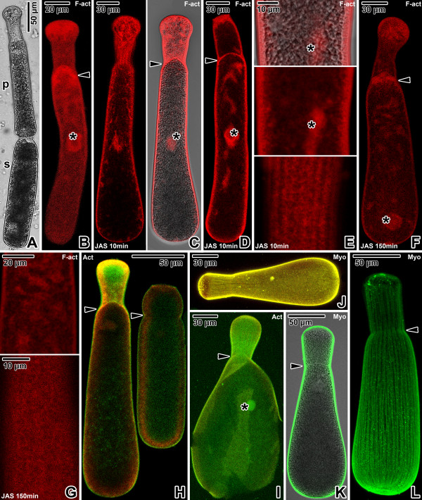
Figure 1.
Actin and myosin in Gregarina cuneata gamonts. A. Gamonts in syzygy; primite (p), satellite (s). LM, transmitted light. B. Localisation of F-actin in a gamont; nucleus (asterisk), septum (arrowhead) between protomerite and deutomerite. CLSM, phalloidin-TRITC. C-D. Localisation of F-actin in gamonts (previously associated in syzygy) treated for 10 minutes with 10 μM JAS. Intense labelling is restricted to the cortex and cytoplasmic F-actin aggregations; septum (arrowhead), nucleus (asterisk). Figure C shows a primite. CLSM (left) and merged CLSM/transmitted light (right), phalloidin-TRITC. Figure D shows a satellite. CLSM, phalloidin-TRITC. E. The deutomerite of a gamont treated for 10 minutes with 10 μM JAS. F-actin localisation corresponds to the cortex and nucleus (asterisk). Upper two figures show the gamont middle plane; lower figure shows the cortex in the area of epicytic folds. Merged CLSM/transmitted light (upper) and CLSM, phalloidin-TRITC. F. F-actin localisation in a gamont treated for 150 minutes with 10 μM JAS. Note the decreased labelling of cell cortex and septum (arrowhead), and formation of numerous cytoplasmic aggregations of F-actin; nucleus (asterisk). CLSM, phalloidin-TRITC. G. The deutomerite of a gamont treated for 150 minutes with 10 μM JAS. F-actin labelling is restricted to the cortex in the area of epicytic folds (lower); cytoplasmic F-actin aggregations (upper). CLSM, phalloidin-TRITC. H. Actin localisation in previously associated gamonts; septum (arrowheads). CLSM, IFA. I. Actin localisation in a gamont ghost; nucleus (asterisk), septum (arrowhead). CLSM, IFA. J. Myosin labelling in a maturing gamont. CLSM, IFA. K. Myosin labelling in a mature gamont is restricted to the cortex, but not to the septum (arrowhead). Merged CLSM/transmitted light, IFA. L. Labelling of myosin in a gamont cortex shows a pattern of longitudinal rows; septum (arrowhead). CLSM, IFA. Figures H, I and J show merged FITC (antibody) and rhodamine (counterstaining with Evans blue) fluorescence channels.

