
Figure 2.
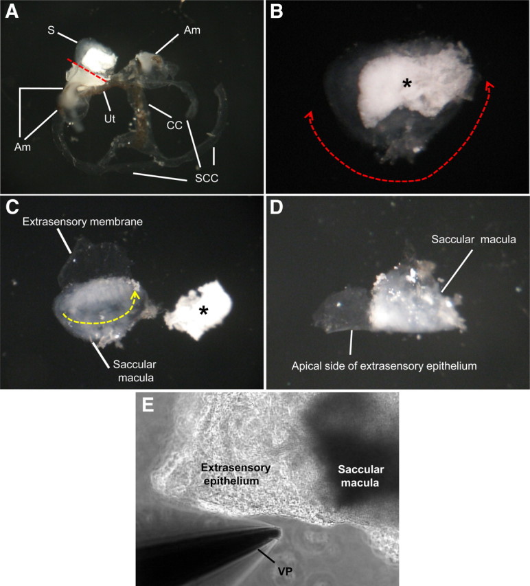
Preparation of saccular extramacular membrane for the measurement of current density. A, Separated membranous labyrinth of vestibule. The saccule is separated with microscissors from the utricle (red dotted line). B, Isolated saccule and separation of extramacular membrane from saccular macula. The extramacular membrane is separated along red dotted line with one side attached to the longest edge of the macula. C, Tissue folding. The tissue is folded along the yellow dotted line with the apical side of the epithelium facing out. D, Prepared tissue before mounting on stage of microscope. E, Photo of mounted tissue for current measurement. S, Saccule; Am, ampulla; Ut, utricle; CC, common crus; SCC, semicircular canal; *otoconia of saccule; VP, vibrating probe.