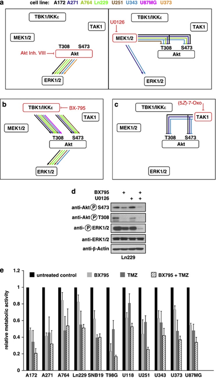
Figure 6.
Noncanonical IKKs activate ERK1/2 via AKT. (a) The indicated cell lines were incubated overnight with the AKT inhibitor VIII (5 μM) or with the MEK1/2 inhibitor U0126 (5 μM), followed by analysis of AKT phosphorylation at T308 or S473 and ERK1/2 phosphorylation by immunoblotting. The results are schematically displayed, the inhibited enzymes are shown in red, the necessity of enzyme activity (as revealed by significantly impaired or absent phosphorylation) is shown by arrows representing the different cell lines. Inhibitory kinase activities (as revealed by exaggerated phosphorylations in the presence of kinase inhibitors) are shown by lines lacking an arrow head. (b) Impact of BX795-mediated TBK1/IKKɛ inhibition and (c) 7-oxozeaenol-mediated TAK1 inhibition on the signaling networks. The original blots for the data sets schematically summarized in (a–c) are shown in Supplementary Figure S8. (d) Ln229 cells were incubated with BX795 (1 μM) and U0126 (5 μM) at the indicated combinations and phosphorylation of ERK1/2 was analyzed by immunoblotting. (e) The indicated GBM cell lines were incubated for 72 h with BX795 (1 μM), temozolomide (TMZ; 100 μM) or a combination of both. Cell viability was determined by MTT assays, error bars show standard deviations from two experiments performed in triplicate.

