Figure 1.
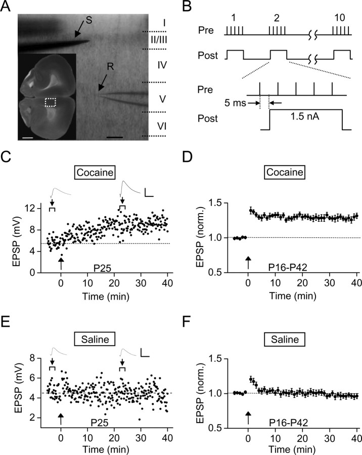
Prenatal cocaine exposure facilitated LTP induction in layer V pyramidal neurons of rat mPFC. A, Images of an acutely isolated coronal slice of P20 rat brain, showing the extracellular stimulating electrode (S) at layer II/III and the whole-cell recording electrode (R) at layer V at the mPFC region (marked by the white box in the inset). Scale bars: box, 1 mm; 200 μm. B, Stimulation protocol for LTP induction (termed mTBS), consisting of presynaptic activation of 10 bursts (each with 5 pulses at 100 Hz) spaced at 200 ms and repeated three times at 10 s intervals and postsynaptic injection of a depolarizing current pulse (1.5 nA, 40 ms) during each burst, with a 5 ms interval between the onset of presynaptic and postsynaptic stimulation. C, An example of LTP induction in a slice prepared from a P25 rat that was exposed to cocaine in utero for 7 d. The data points represent the amplitude of EPSPs recorded before and after application of mTBS (at the time marked by arrow). Sample traces above represent averages of 10 EPSPs at the marked time (arrowhead). Calibration: 6 mV, 50 ms. D, Summary of data from all experiments similar to that in C, showing normalized EPSP amplitudes before and after LTP induction in slices from P16–P42 rats that were prenatally exposed to cocaine (for 7 d from E15 to E21; n = 40). The EPSP amplitudes over 1 min intervals were normalized by the mean amplitude before induction. Error bars indicate SEM. E, F, Same as C and D except that the results were obtained from slices of prenatal saline-treated rats (F) (n = 21).

