
Figure 7.
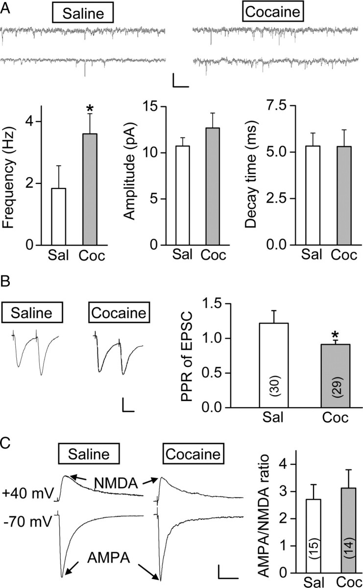
Prenatal cocaine exposure enhanced excitatory glutamatergic transmission of mPFC layer V pyramidal neurons. A, Comparison of the frequency, averaged amplitude, and decay time of mEPSCs from P23–P26 rats prenatally treated with cocaine or saline, with two sample recordings of mEPSCs for each group shown above (n = 12 for each group; *p = 0.016, t test). Calibration: 10 pA, 20 s. B, Left, Sample traces of paired EPSCs (average of 15) in response to two sequential presynaptic stimuli at an interval of 50 ms. Calibration: 100 pA, 25 ms. Right, Summary of PPR (EPSC amplitude of the second divided by the first). Data were obtained from P23–P26 rats prenatally treated with cocaine or saline. Number associated with the histogram refers to the total number of cells recorded. *p = 0.034, t test. C, Left, Sample traces of glutamatergic currents mediated by AMPA and NMDA receptors, recorded at −70 and +40 mV, respectively. Recordings were from P25–P26 rats prenatally treated with saline or cocaine. Calibration: 50 pA, 50 ms. Right, Average AMPA/NMDA ratio obtained by dividing the peak amplitude of AMPA and NMDA receptor-mediated EPSCs. Data were obtained from P24–P26 rats. Number associated with the histogram refers to the total number of cells recorded. Error bars indicate SEM.