
Figure 8.
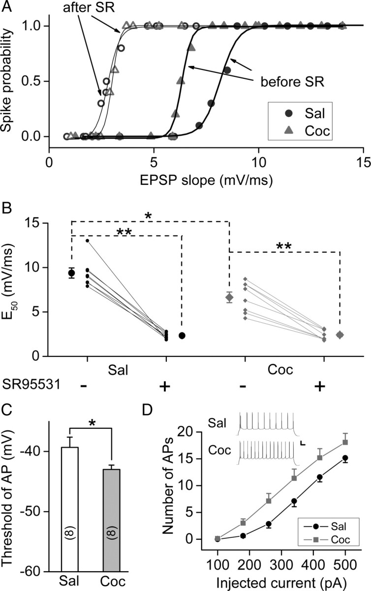
Prenatal cocaine exposure increased the E–S coupling and excitability of mPFC layer V pyramidal neurons. A, E–S coupling studies on rats prenatally exposed to saline (Sal) or cocaine (Coc) in the absence (“before”) and presence (“after”) of SR95531 (SR) (0.5 μm). The curves represent the best fit with the sigmoidal function. B, The values of E50 before and after bath-application of SR95531 (0.5 μm). E50 represents the efficacy of E–S coupling, as defined by the EPSP slope that initiates spiking with a probability of 0.5. Data obtained from the same neuron were connected by the line. Recording were performed in the slices obtained from P24–P27 rats. *p < 0.05, **p < 0.01, t test. C, Summary of the threshold potential for AP initiation. The threshold was determined by the membrane potential at the peak of the maximal subthreshold EPSP. *p < 0.05, t test; n = 9; the same data set as in B. D, Changes of the excitability of mPFC layer V pyramidal neurons resulting from prenatal cocaine exposure, as shown by the number of APs triggered by injection of depolarizing currents of various amplitudes (duration, 800 ms). Data were obtained from the recording on the slices obtained from P19–P27 rats. Inset, Examples of APs elicited by a constant depolarizing current (500 pA, 800 ms) in mPFC neurons from prenatal saline- and cocaine-treated P25 rats (n = 17–24). Calibration: 20 mV, 80 ms. Error bars indicate SEM.