
Figure 1.
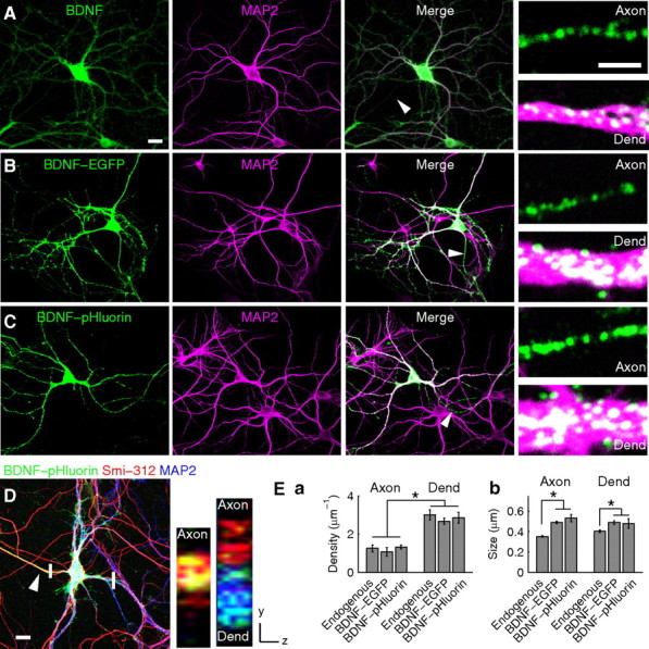
Expression and localization of endogenous BDNF and BDNF–EGFP/pHluorin in cultured hippocampal neurons. A, Coimmunostaining of endogenous BDNF (green) and MAP2 (magenta) in cultured hippocampal neurons. Arrowhead, Axon. Small panels, Higher-magnification images of an axon and a dendrite (Dend). Scale bars: left, 20 μm; small panels, 2 μm. B, C, Neurons expressing either BDNF–EGFP (B) or BDNF–pHluorin (C) were immunostained for EGFP (green) and MAP2 (magenta). Arrowheads, Axons. Small panels, Higher magnification of axons and dendrites. Scale bars are the same as in A. D, A neuron triple immunostained with antibodies to EGFP (green), axon (arrowhead)-specific Smi-312 (red), and MAP2 (blue). Small panels, Reconstructed images along the axial plane (at regions marked by white bars) of a single BDNF–pHluorin-expressing axon (left) and of an axon not expressing BDNF–pHluorin (right) together with a BDNF–pHluorin-expressing dendrite (right). Scale bar, 20 μm. E, Bar graphs showing the density (Ea) and size (Eb) of BDNF-containing fluorescence puncta. *p < 0.01, significant difference between axon and dendrite by two-way ANOVA and t test.