Figure 8.

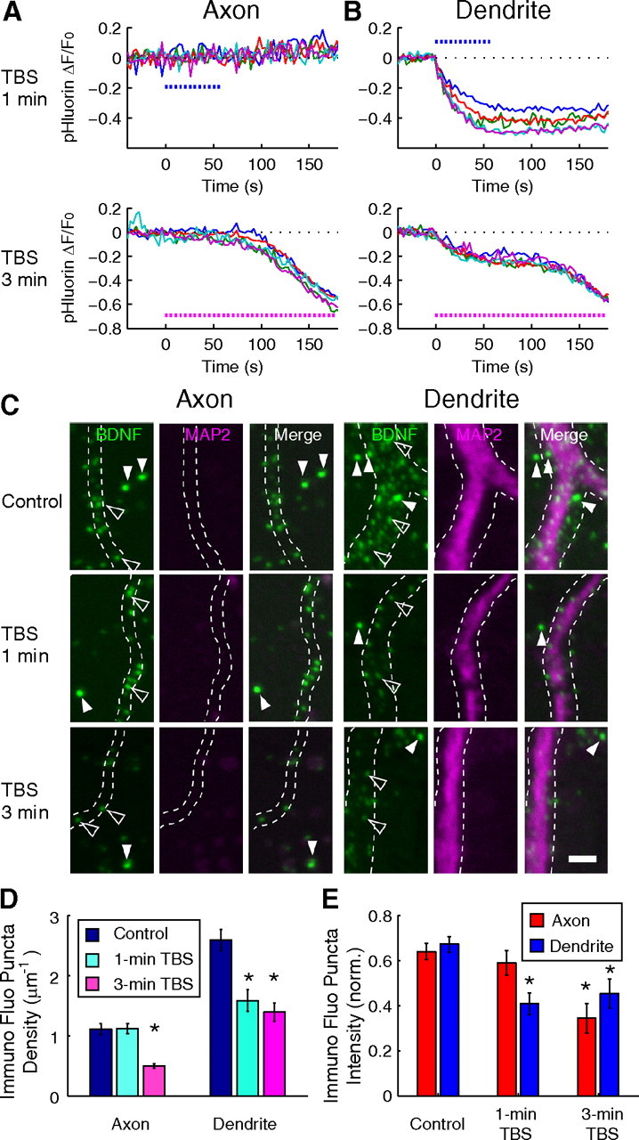
Endogenous BDNF exhibits similar activity-induced secretion as BDNF–pHluorin. A, B, Sample traces of fluorescence changes at individual BDNF–pHluorin puncta induced by 1 or 3 min TBS at axon (A) and dendrite (B), before immunostaining for endogenous BDNF. C, Neurons in the same culture as those in A and B were immunostained for endogenous BDNF immediately after 1 or 3 min TBS. Open arrowheads, Puncta of immunostained endogenous BDNF. Filled arrowheads, Green fluorescent beads added to the culture after immunostaining for normalization. Scale bar, 2 μm. D, E, Bar graphs showing the density of endogenous immunostained BDNF puncta (D) and the intensity of endogenous BDNF puncta fluorescence, normalized by that of the microspheres (E), with or without exposure to 1 or 3 min TBS, for axons (n = 5 neurons) and dendrites (n = 5 neurons). Error bars indicate SEM.
