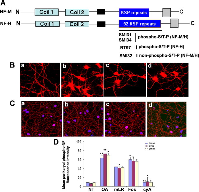
Figure 1.
Effect of protein phosphatase inhibitors on perikaryal phosphorylation of NF. A, Structure of rat NF-M/H representing KSP repeats in the tail domain. The KSP repeats are recognized by phospho-NF-M/H antibodies (SMI31, SMI34) and phospho-NF-H antibodies (RT97). The SMI32 antibodies recognize the non-phospho-KSP repeat region of NF-M/H. B, The dissociated E18 rat primary cortical neurons were cultured for 7 d in situ and then treated with or without different protein phosphatase PP2A inhibitors for 2.5 h [OA (b; 0.25 μm), microcystin LR (c; 15 μm), or Fos (d; 1 μm)] and then subjected to immunofluorescence with phospho-NF (SMI31) antibodies. Control (a) shows phosphorylated NF in processes with no phosphorylation in cell bodies. There was a significant increase of somatic phosphorylation in OA-treated (b), mLR-treated (c), and fostriecin-treated (d) neurons. C, The 7 DIC cortical neurons were subjected to the dose-dependent treatment of the PP2B-specific inhibitor cyclosporine A [0.5 μm (b), 1 μm (c), and 2.5 μm (d)]. Scale bar, 10 μm. D, Densitometry analyses of phospho-NF was performed with images captured using NIH Image software. To determine the relative intensity of SMI31 immunoreactivity within perikarya, representative areas of perikarya excluding obvious vesicles and the nucleus were quantified using the freehand selection tool. Similar results were obtained using RT97 and SMI34 antibodies. Intensity of SMI31 (blue), RT97 (red), and SMI34 (yellow) immunoreactivity (p-NF-H) in neuronal cell bodies was analyzed with the NIH ImageJ histogram from 50 to 100 individual cells from at least three experiments. The mean signal intensity (total pixel density per number pixels) is shown. Values in graphs are means ± fluorescence intensity (in arbitrary densitometric units) in perikarya from three different experiments. Mean fluorescence of phosphorylated NF-M/H immunoreactivity. *p < 0.01 and **p < 0.001 of phospho-NF-M/H in perikarya relative to fluorescence intensity values of nontreated neurons (NT).

