
Figure 6.
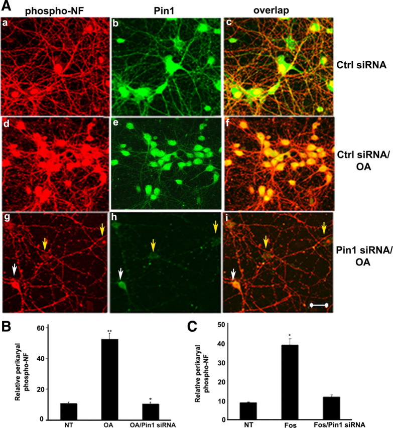
Knockdown of Pin1 by Pin1 siRNA inhibits OA-induced perikaryal phosphorylation of NF-M/H. A, Five DIC cortical neurons were transfected with either control (Ctrl) and scrambled siRNA (a–f) or Pin1 siRNA (g–i) and then treated with 0.25 μm OA (d–i) on day 7 for 2.5 h. Neurons were immunostained, p-NF-H was detected using SMI31 (red), and Pin1 was detected by using Pin1 antibody (green). Neurons transfected with Pin1 siRNA exhibited reduced p-NF-H in the cell body. Nontreated neurons exhibited SMI31 staining in the processes with little or no staining in the cell body (a–c), which is increased during OA treatment (d–f). Cell body SMI31 staining was reduced in Pin1 siRNA-treated neurons (g–i). Scale bar, 20 μm. B, Representative neurons are treated with OA as indicated. Intensity of SMI31 immunoreactivity (phospho-NF) in neuronal cell bodies was analyzed with the NIH ImageJ histogram. The mean signal intensity (total pixel density per number pixels) is shown. Values in graphs are means ± fluorescence intensity (in arbitrary densitometric units) in perikarya from four different experiments. Note the accumulation of phospho-NF in perikarya in OA-treated neurons and restoration of normal levels of p-NF-H in Pin1 siRNA-treated cortical neurons. C, Intensity of SMI31 immunoreactivity (phospho-NF) in neuronal cell bodies was analyzed with the NIH ImageJ histogram after treatment of cortical neurons with Fos. *p < 0.01, phospho-NF-M/H in perikarya relative to fluorescence intensity values of control. NT, Nontreated.