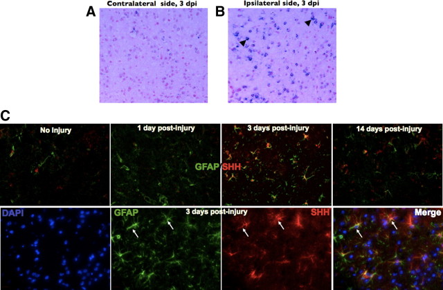
Figure 2.
SHH is expressed in the mouse brain after brain injury. A, B, In situ hybridization for SHH in the uninjured contralateral hemisphere and injured brain, respectively. Black arrowheads indicate positively stained cells (blue) in the injured right cortex. C, Top, from left to right, SHH and GFAP double immunofluorescence from uninjured brain and brains processed after 1, 3, and 14 d after injury, respectively. C, Bottom, from left to right, High-magnification immunofluorescence staining of a mouse brain at 3 d after injuring showing 4′,6′-diamidino-2-phenylindole (DAPI), GFAP, SHH, and a merged image, respectively. White arrows indicate astrocyte costaining for SHH and GFAP. Images in A, B, and C (top) were taken at 20× magnification. Images in C (bottom) were taken at 63× magnification. dpi, Days post-injury.

