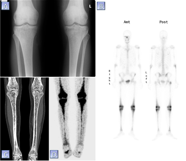
Figure 1.
Diagnostic imaging in ECD. Various modalities in the skeletal assessment of a single ECD patient. (A) A plain radiograph of the knees demonstrating bilateral sclerotic changes in the femoral and tibial bones. (B) 99mTc bone scintigraph taken prior to the diagnosis of ECD. Note the abnormally increased tracer uptake especially involving the periarticular region of the femurs and the tibiae. (C) Coronal reconstruction of a computed tomography study of the femurs and tibiae. Note the diffuse, irregular intra-medullary lytic-sclerotic pattern as well as the marked cortical thickening of the tibiae. (D) Coronal reconstruction of a positron emission tomography taken for the purpose of follow up 4.5 years pursuant the diagnosis of ECD. This study shows bilateral symmetric abnormally increased intra-medullary uptake of fluorodeoxyglucose in the femurs and tibiae.

