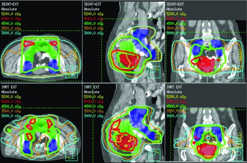
Figure 1.

Axial, sagittal, and coronal images for 3D-CRT (top panels) and IMRT (bottom panels) plans in a patient with T4 disease. The primary tumor (shaded red) received 50 Gy, and the internal iliac, mesorectal, and presacral nodes (green) and the external iliac nodes (blue) received 45 Gy. IMRT improved target coverage, while limiting dose to surrounding normal organs, including the small bowel.
