Figure 4.
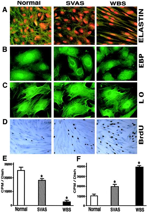
A, Representative photomicrographs of 10-d-old cultures of human aortic SMC immunostained with specific antibodies to elastin (magnification ×100). Nuclei were counterstained with propidium iodide. B and C, Representative higher-power images (magnification ×400) comparing expression of the EBP and LO in 3-d-old cultures of aortic SMC. D, Representative photomicrographs of 10-d-old cultures of human aortic SMC metabolically labeled with BrdU and immunostained with anti-BrdU antibodies (brown nuclei), demonstrating higher-than-normal proliferating rates of SVAS cells and of WBS cells (magnification ×40). E and F, Results (mean ± SD) of quantitative analysis of a typical experiment using 3-d-long metabolic labeling of quadruplicate cultures with radioactive valine, followed by biochemical isolation of insoluble elastin, which demonstrates that aortic SMC from patients with SVAS and patients with WBS deposit, respectively, low and very low amounts of insoluble elastin (E), and of measurements of [3H]-thymidine incorporation into the parallel quadruplicate cultures, which indicate that abnormally low levels of insoluble elastin are associated with an inversely proportional increase in the proliferation rate of SMC in patients with SVAS and patients with WBS (F) (* [P<.001]).

