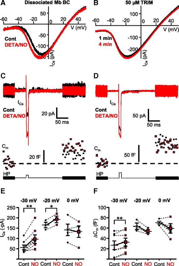
Figure 6.

The NO donor-mediated shift of ICa caused weighted potentiation of Mb output selectively in response to weak stimuli. A, Application of NO donor DETA/NO (1 mm) for 1.5 min shifted the ramp-evoked ICa activation to more negative potentials in enzymatically dissociated Mbs (black represents control; red represents DETA/NO). B, Inhibition of endogenous NO synthases by TRIM (50 μm) prevented the leftward shift of ICa activation in axotomized Mb terminals in slice preparation during consecutive ramp stimulations (black represents control; red represents second ramp I-V). C, Bath application of DETA/NO (1 mm) facilitated ICa and enhanced exocytosis (Cm) from the axotomized Mb terminals in response to a depolarizing step from −60 mV to −30 mV. HP, Holding potential. Black represents control; red represents DETA/NO treatment. D, Bath application of DETA/NO (1 mm) slightly increased ICa, but this increase was not associated with increased exocytosis from axotomized Mb terminals in response to a depolarizing step from −60 mV to −20 mV (bottom trace). Black represents control, red represents DETA/NO treatment. E, Summary figure displaying DETA/NO (1 mm) effect on peak ICa in response to 10 ms step from −60 mV to −30, −20, or 0 mV. Black circles represent control; red squares represent DETA/NO. **p = 0.004 (paired Student's t test). n = 7. *p = 0.008 (paired Student's t test). n = 3. F, Summary figure displaying DETA/NO (1 mm) effect on ΔCm evoked by 10 ms step from −60 mV to −30, −20, or 0 mV. Black circles represent control; red squares represent DETA/NO. **p = 0.003 (paired Student's t test). n = 7. n = 3 for −20 mV; n = 4 for 0 mV. Every terminal was tested at one depolarization level in control then in the presence of DETA/NO; thus, each pair of measurements shown originated from different cells. Data are mean ± SEM.
