
Figure 6.
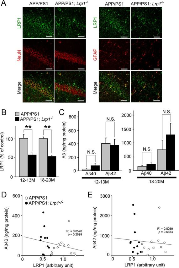
LRP1 deletion in neurons in APP/PS1 mice does not affect Aβ levels in the hippocampus. A, Hippocampus from control APP/PS1 and APP/PS1; Lrp1−/− mice were costained with an LRP1 antibody and a neuronal marker NeuN or an astrocyte marker GFAP at 12 months of age. Scale bar, 50 μm. B, LRP1 expression in the cortex from APP/PS1 and APP/PS1; Lrp1−/− mice was detected by Western blot at 12–13 (n = 6–7) and 18–20 months (n = 5–6) of age. C, The concentrations of insoluble Aβ40 and Aβ42 levels in the hippocampus from control APP/PS1 and APP/PS1; Lrp1−/− mice analyzed by ELISA at 12–13 (n = 6–7) and 18–20 months (n = 5–6) of age. Data were plotted as mean ± SEM; **p < 0.01. N.S., Not significant. D, E, The correlations of insoluble Aβ40 (D) and Aβ42 (E) levels with LRP1 levels in the hippocampus are plotted. APP/PS1 (n = 12) and APP/PS1; Lrp1−/− mice (n = 11) were analyzed at 12–20 months of age. LRP1 levels were quantified by Western blot and normalized to β-actin. Data were plotted as the ratios to mean value of control APP/PS1 mice. The correlation coefficient (R2) and p values are shown in the graph.