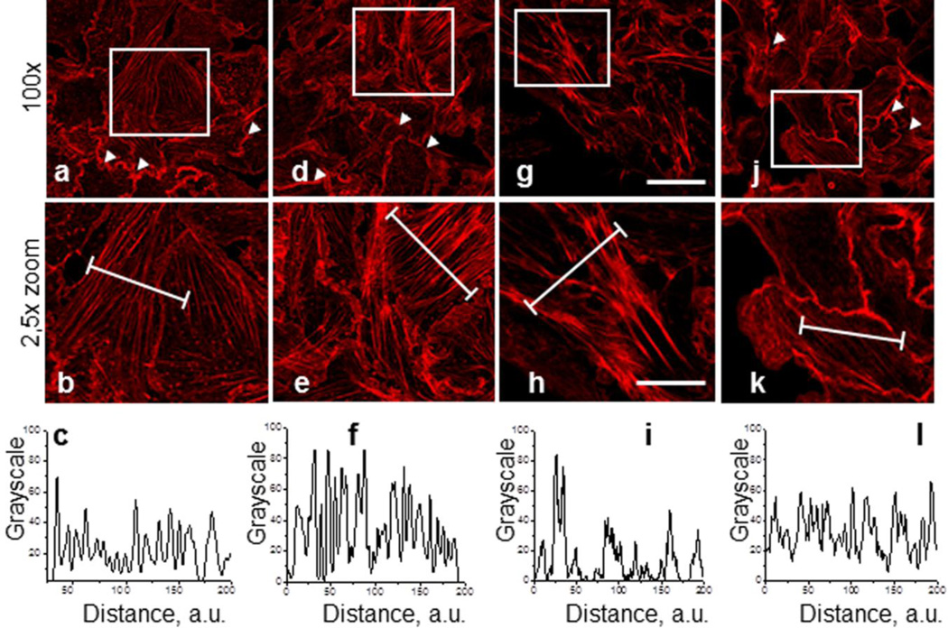
Fig. 1.
Effect of the Arp2/3 complex inhibition with CK-666 and CK-689 on actin cytoskeleton organization in M-1 epithelial cells. M-1 cells were pretreated with vehicle (a) CK-0499666 (CK-666) in concentrations of 100 μM and 200 μM for 2 hrs (d), and g, respectively, or CK-689 (j, 2 hrs, 200 μM). Corresponding close-up images are shown under full images (b, e, h, and k respectively). Images were taken from M-1 cells stained with rhodamine-phalloidin to visualize actin microfilaments. Scale bar is common for all the images of the row and is 25 and 10 μm for upper and lower rows, respectively. Bottom row (c, f, I, l) demonstrates relative fluorescence across a region of interest (white lines in correcponding close-up images).

