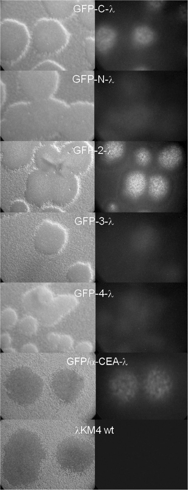
Figure 3.

Microscope observation of the lambda phage plaques displaying GFP as a fusion to the different sites of the gpD protein. Lambda phage clones displaying GFP as C-terminal (GFP-C-λ), N-terminal (GFP-N-λ) fusion, fused to the new positions inside of the gpD protein (GFP-2-λ, GFP-3-λ, GFP-4-λ) or double displaying GFP/α-CEA-λ, were observed in fluorescent and light microscope. λKM4 wild type phage is included in the analysis as a negative control.
