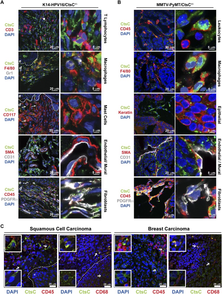
Figure 2.
Stromal and epithelial cells express CtsC. Localization of CtsC (green) within dysplastic skin of HPV16/CtsC+/− mice (6 mo of age) (A) and mammary tumors from MMTV-PyMT mice (B). CtsC was highly expressed in F4/80+ macrophages, CD45−PDGFRα+ fibroblasts, and epithelial cells in both transgenic models. While CtsC was also expressed by CD31+ endothelial cells and SMA+ pericytes in mammary tumors, CtsC localization was prominent in CD117+ mast cells in HPV16/CtsC+/− ear tissue. (C) Immunofluorescent confocal microscopy of human skin SCCs and breast tumors revealing CtsC expression (green) in the stromal compartment, which colocalized to infiltrating CD45+ immune cells and CD68+ macrophages (red), in addition to localization to the epithelial compartment of tumors. (Dashed line) Epithelial–dermal interface; (e) epidermis.

