Figure 1.

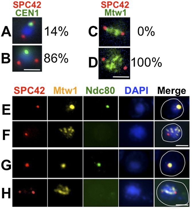
Kinetochore disassembly blocks precocious kinetochore–microtubule interactions. (A) ipl1-as5/ipl1-mn cells (DSY434 and DSY436) were sporulated. 1-NA-PP1 was added (T = 5 h) to allow spindle formation during the pachytene arrest (no β-estradiol added). The distance between CEN1 (CEN1 was tagged with GFP using the lacO/GFP-lacI system) (Straight et al. 1996; Obeso and Dawson 2010) and the closer SPB (Spc42-DsRed) was measured. (A) CEN1 at the pole (≤0.5 μm from the nearest pole). (B) CEN1 distant from the pole (>0.5 μm from the nearest pole) (n = 84). (C,D) ipl1-as5/ipl1-mn cells (DSY428) were sporulated, and 1-NA-PP1 was added (T = 5 h) to allow spindle formation during pachytene arrest (ndt80Δ). Kinetochores (Mtw1-GFP) and SPBs (Spc42-DSRed) were visualized. Examples of a wild-type cell with chromosomes aligned on the metaphase spindle (C) and an ipl1 cell with scattered chromosomes on a precocious spindle (D). In a sample of 44 ipl1 cells (at T = 9 h), 100% had scattered chromosomes. (E–H) ndt80Δ (DSY585) (E,F) and ndt80Δ ipl1-as5/ipl1-mn (DSY588, 1-NA-PP1 at 5 h) (G,H). Samples were prepared for whole-cell immunostaining for kinetochores (Mtw1-myc and Ndc80-GFP) and SPBs (Spc42-DsRed) at meiotic entry (E,G) and prophase (F,H). (E) Clustered Ndc80 was observed in 98% of the cells at meiotic entry (marked by clustered Mtw1; n = 55). (F) Ndc80 was absent in 100% of prophase cells (marked by dispersed Mtw1; n = 31). (G) Clustered Ndc80 was observed in 100% of cells at meiotic entry (marked by clustered Mtw1; n = 51). (H) Ndc80 was absent in 100% of prophase cells (marked by dispersed Mtw1; n = 31). Bar, 2 μm.
