Figure 4.

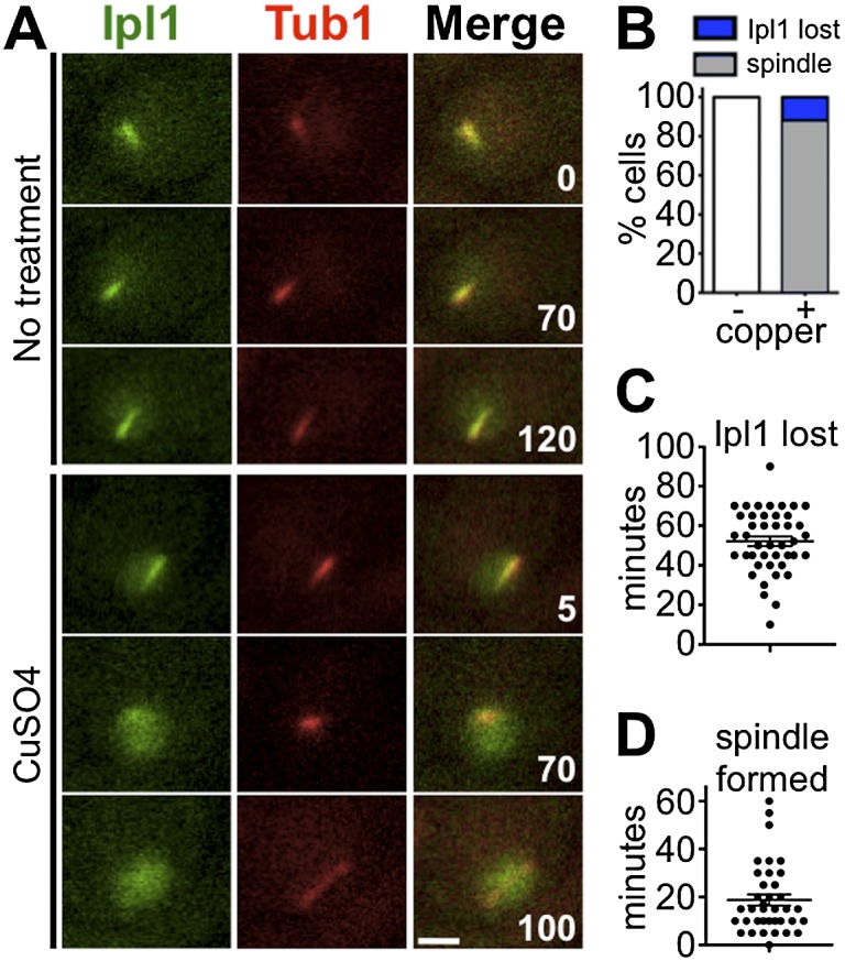
Ipl1 delocalizes from the prophase microtubule array before spindle formation. (A–D) Ipl1 localization (IPL1-EGFP) relative to the prophase microtubule array (mCherry-TUB1) was monitored after induced expression of CLB4 (PCUP1-CLB4) while in pachytene-arrested cells (ndt80Δ) (DHC244 and DHC245). (A) Meiosis was induced. At T = 4 h, cells were transferred to a microfluidic plate. Images were collected every 5 min for 120 min. After the first frame, sporulation medium with or without CuSO4 was introduced into the chamber. (B) Cells were scored for delocalization of Ipl1 from the SPBs and formation of spindles. All cells that formed spindles also lost SPB localization of Ipl1. (C) Cells were scored for the number of minutes after copper addition at which Ipl1 was lost from the SPBs. (D) Cells were scored for the number of minutes after Ipl1 delocalization at which a spindle formed. n > 40 for each sample. Bar, 2 μm.
