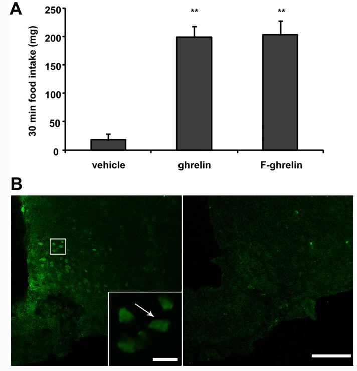
Figure 1. F-ghrelin increases food intake and specifically binds to brain areas in mice.
Panel A shows 30 min food intake in mice ICV injected with vehicle alone or containing 16.6 pmol of ghrelin or F-ghrelin. Data represent the mean ±SEM. **, p ≤ 0.01. Panel B shows confocal representative photomicrographs of fluorescein-IR signal in ARC coronal sections of mice that were ICV injected with 16.6 pmol of F-ghrelin alone (left) or plus 166 pmol of ghrelin (right). Insert shows high magnification of the area marked in the low magnification image. Arrows point fluorescein-IR cells. Scale bars, 100 μm (low magnification), 10 μm (high magnification).

