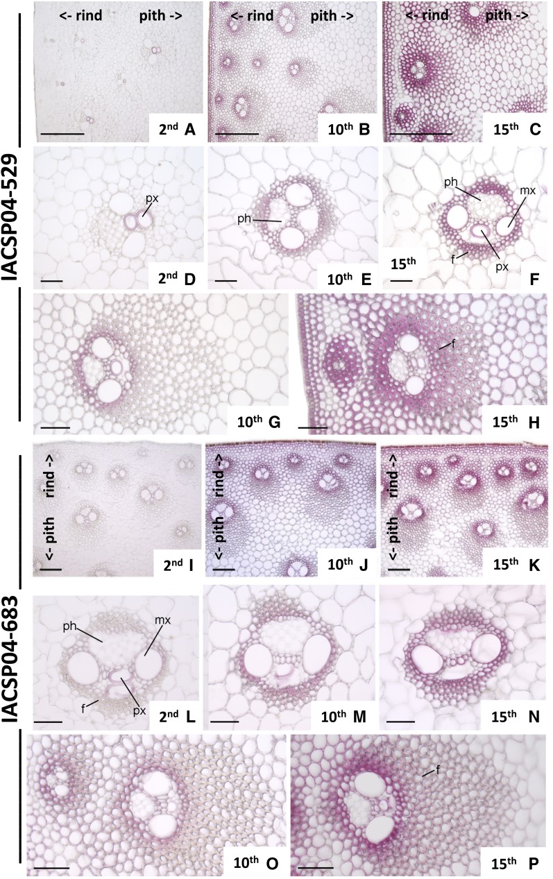
Figure 2.
Transverse sections of internodes of two sugarcane genotypes stained for lignin with phloroglucinol. Genotypes IACSP04-529 (A–H) and IACSP04-683 (I–P) are shown. Details are as follows: second internode (A, D, I, and L); 10th internode (B, E, G, J, M, and O); 15th internode (C, F, H, K, N, and P); peripheral region (A–C and I–K); detail of the vascular bundle of the central region (D–F and L–N); and detail of the vascular bundle of the peripheral region (G, H, O, and P). f, Fibers; mx, metaxylem; ph, phloem; px, protoxylem. Numbers indicate the positions of the internode. <- rind and pith-> indicate their positions in the photographs. Bars = 200 μm (A–C) and 50 μm (D–M).

