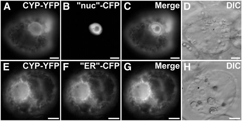
Figure 6.
CYP71D351 (T16H2) is located to the ER. C. roseus cells were transiently transformed with CYP71D351-YFP-expressing vector (CYP-YFP; A and E) in combination with plasmids expressing a nucleus-CFP marker (“nuc”-CFP; B) or an ER-CFP marker (“ER”-CFP; F). Colocalization of the two fluorescence signals appears on the merged images (C and G). Cell morphology (D and H) was observed with differential interference contrast (DIC). Bars = 10 µm.

