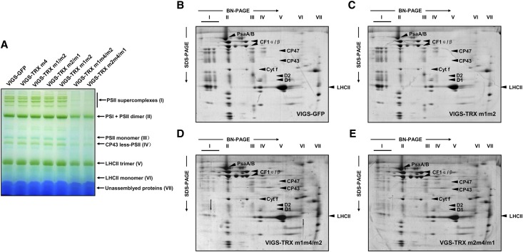
Figure 4.
Analyses of thylakoid protein complexes from VIGS plants. A, BN-PAGE analyses of thylakoid chlorophyll-protein complexes. Equal amounts of thylakoid membranes (8 µg of chlorophyll) from VIGS-TRX m and VIGS-GFP plants were solubilized with 1% (w/v) DM and separated by BN-PAGE. Assignments of the thylakoid membrane macromolecular protein complexes indicated at right were identified according to a previous study (Peng et al., 2006). B to E, Two-dimensional BN/SDS-PAGE fractionation of thylakoid protein complexes. Individual lanes from the BN-PAGE gels in A were subjected to second-dimension SDS-urea-PAGE followed by Coomassie blue staining. Identities of the relevant proteins are indicated by arrows. Three biological replicates were performed, and similar results were obtained. [See online article for color version of this figure.]

