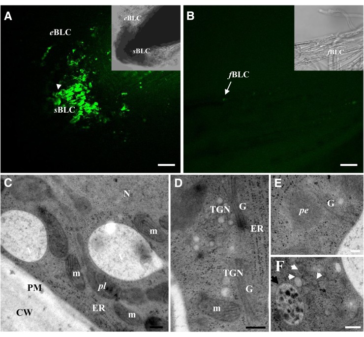
Figure 2.
Root border-like cells of flax are released as living cells. A and B, Root border-like cells stained with calcein-AM. The presence of fluorescence is indicative of the cell viability. Note the presence of fluorescence in spherical border-like cells and elongated border-like cells in A. No fluorescence is observed in filamentous border-like cells. C to F, Ultrastructural organization of spherical border-like cells. Cytoplasm showing different organelles, including Golgi stacks (G), endoplasmic reticulum (ER), mitochondria (m), and nucleus (N). White arrowheads indicate secretory vesicles, and the black arrowhead shows a multivesicular body. CW, Cell wall; eBLC, elongated border-like cells; fBLC, filamentous border-like cells; pe, peroxisome; pl, plastid; PM, plasma membrane; sBLC, spherical border-like cells; TGN, trans-Golgi network. Bars = 40 μm (A and B) and 200 nm (D–F). [See online article for color version of this figure.]

