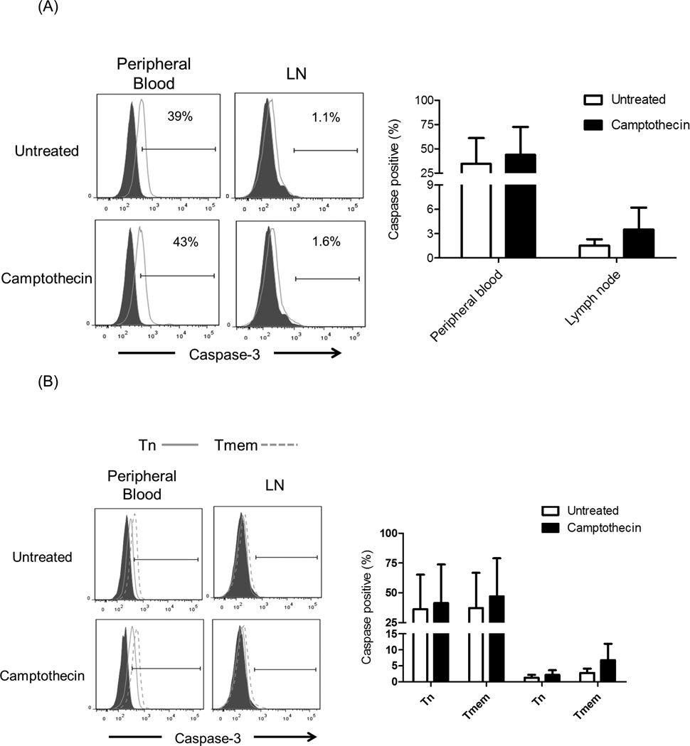
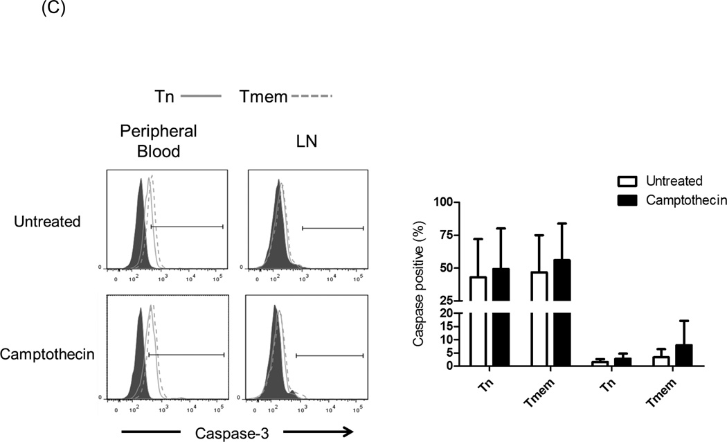
Fig. 6.
Caspase-3 expression. Cynomolgus monkey peripheral blood (PB) and lymph node (LN) cells were incubated with 5µM Camptothecin for 8 hours. Control samples were untreated cells. Following culture, the cells were stained for intracellular expression of Caspase-3 by flow cytometry. Caspase-3 expression in (A) CD3+T cells, (B) CD4+T cells, and (C) CD8+T cells was evaluated. Histograms (left) are from one representative monkey from each group; grey histograms indicate isotype controls. Graphs (right) represent mean values obtained from normal monkeys (n=4).


