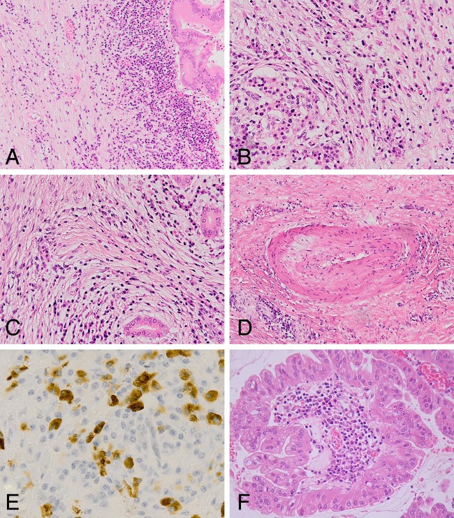
Figure 1.
Histological appearances of case 1 (A–E) and case 14 (F). (A) Medium power view showing edge of intraduct papillary mucinous neoplasm (IPMN) with chronic inflammation and fibrosis extending into the adjacent pancreas. (B) Lymphoplasmacytic inflammation adjacent to a surviving islet of Langerhans within the distant pancreas. (C) Storiform fibrosis. (D) Obliterative venulitis. (E) IgG4 immunohistochemistry revealing numerous IgG4+ plasma cells within the inflammatory infiltrate. (F) Oncocytic variant of IPMN showing prominent lymphoplasmacytic inflammation within a papillary core.

