Fig. 3.
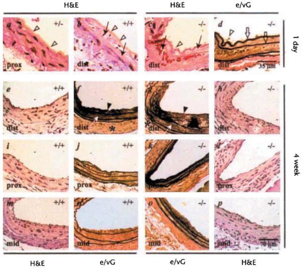
Vascular hyperplasia following carotid injury occurs in Fgf2+/+ mice. Carotid arteries were injured in adult mice and examined for the development of hyperplastic vascular layers. a–d, Hematoxylin and eosin (H&E)-stained sections from vessels 1 day after injury from control (Fgf2+/+, Fgf2+/+ n = 4) and Fgf2−/− (n = 5) mice show a range of injury features from (a) endothelial cell denudation with platelet deposition (open arrowheads) proximal to the heart (prox) to (b) severe medial damage leading to medial hemorrhage (arrow) distal to the heart near the thyroid (dist). c, H&E-stained section of an Fgf2−/− mouse carotid showing severe medial injury, d, Serial section of c stained with an elastin/van Gieson stain (e/VG) to highlight the elastic layers, which remained intact (open arrows), e–p H&E- and e/VG-stained serial, section pairs from 4-week post-injury carotids from Fgf2−/− (n = 3) and Fgf2+/+ (n = 2) mice show hyperplastic responses involving the adventitial (*), medial (white arrow), and intimal (closed arrow heads) layers. Panels e, f, i, and j are from the same wild-type mouse showing lesions from two positions (dist and prox). Panels g, h, k, and l show lesions from the same Fgf2 knockout mouse. Panels m, n, o, and p show lesions at the middle position (mid) along the injured carotid from a wild-type (m, n) and Fgf2−/− mouse (o, p). The scale bar shown in d applies to all 1-day panels; the dimension scale in p is the same for all 4-week panels.

