
Fig. 2.
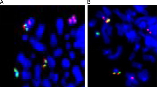
Fluorescent in situ hybridization. (A) FISH on partial metaphase with t(9;22)(q34;q11) using BCR/ABL Dual Color Dual Fusion translocation, Vysis(D-FISH) and Human Chromosome 9 Centromeric Cambio probe (20 metaphases and 200 cells analyzed). D-FISH probe: fusion signal (yellow) was present on Philadelphia chromosome and on the long arm of derivative chromosome 9, one orange signal (ABL probe) was present on the long arm of normal chromosome 9, and one green signal (BCR probe) was present on the long arm of normal chromosome 22. Human Chromosome 9 Centromeric Cambio probe: two red signals were present on normal and derivative chromosome 9. (B) FISH on partial metaphase with t(11;22)(p15;q11) using BCR/ABL Dual Color Dual Fusion translocation, Vysis (D-FISH) and Human Chromosome 11 Centromeric Cambio probe (20 metaphases and 200 cells analyzed). D-FISH probe: fusion signal (yellow) was present on der(22) and on the short arm of derivative chromosome 11, one orange signal (ABL probe) was present on the long arm of normal chromosome 9, and one green signal (BCR probe) was present on the long arm of normal chromosome 22. Human Chromosome 11 Centromeric Cambio probe: two red signals were present on normal and derivative chromosome 11. (For interpretation of the references to color in this figure legend, the reader is referred to the web version of this article.)