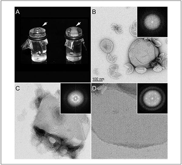
Figure 17.15.4.

Examples of a two-dimensional crystallization assays. (A) A picture of two dialysis buttons with a well containing the protein-lipid-detergent mixture (arrows) sealed with a dialysis membrane. The button on the left is at the start of the experiment while the button on the right represents the end point. As vesicles begin to form, they scatter light, resulting in a translucent whitish/gray solution inside the well (arrow). The buttons are then opened and the solution prepared for analysis by negative-stain EM as presented in panels B-D. Proteins incorporated in to the lipid bilayer can adopt multiple conformations, with the most common being amorphous (B), small mosaic crystals (C), and large well-ordered 2D crystals (D). Inset panels are the corresponding Fourier transforms. Noncrystalline amorphous structures do not show reflections in Fourier transforms (A), while vesicles containing crystalline protein will produce reflections (C and D). Mosaic crystals (C) will lead to a Fourier transform with overlapping reflections that appear as a ring caused by multiple overlapping lattices. Well-ordered crystals as presented in (D) that produce strong and sharp reflections in Fourier transforms are ready for analysis by cryo EM for high-resolution 3D structure determination.
