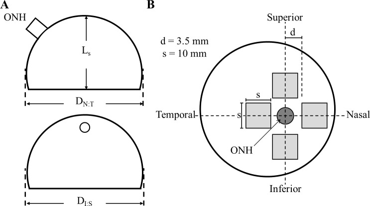
Figure 1.
(A) Relevant physiological measurements of the posterior sclera taken before dissection. D is the average of the globe diameters DN:T and DI:S, and Ls is the specimen axial length in the anterior to posterior direction. (B) Regional sections which were 1.0 cm2 obtained for SALS measurement are shown in light gray; d is the distance from the center of the optic nerve to the start of the section, which was 3.5 mm on average. Our samples were therefore obtained from a region approximately 2.25 mm from the edge of the optic nerve.

