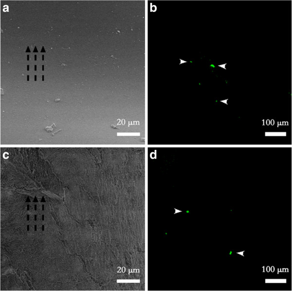
Figure 4.

Low vacuum SEM and immunofluorescent image of platelet adhesion on vascular grafts with different topography. (a) Nothing but small amount of cellular debris adhered on the smooth topography. (c) Protein-like substances adhered on the aligned topography along the direction of blood flow and aligned nanofibers. (b) ~ (d) Few platelets adhered on both topography. Broken black arrows indicate the direction of the blood flow, and white arrows indicate the adhered platelets.
