
Figure 1.
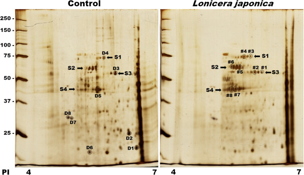
Two-dimensional electrophoresis maps of control and Lonicera japonica-photosensitized CH27 cells. Cells were incubated with 0.1% DMSO or 100 μg/ml Lonicera japonica extracts for 4 h and then irradiated with 0.8 J/cm2 fluence dose. Proteins were separated on a pH 4-7 IPG-strip (7 cm) in the first dimension and on a 12% SDS-polyacrylamide gel in the second dimension. Staining of the protein spots was accomplished by silver nitrate. Results are representative of three independent experiments.