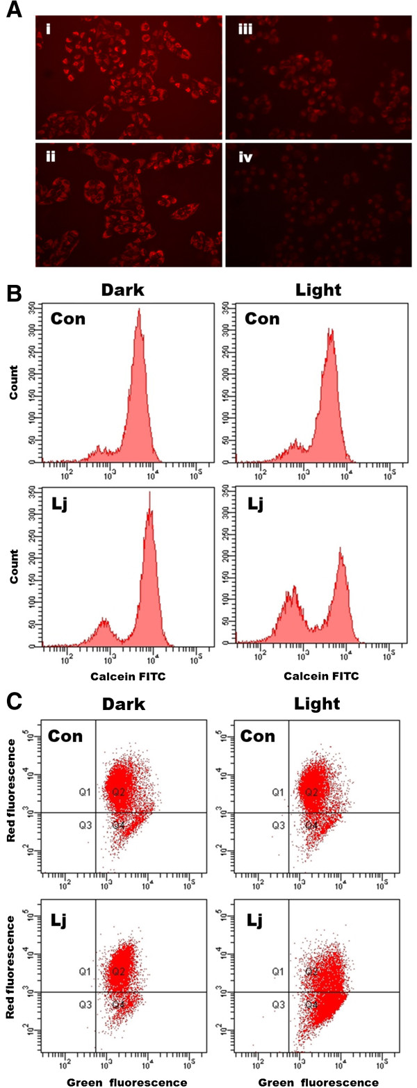
Figure 2.

Effects of photoactivated Lonicera japonica extract on mitochondrial function in CH27 cells. (A) Effects of photoactivated Lonicera japonica extracts on the activity of mitochondria in CH27 cells. CH27 cells were incubated with 0.1% DMSO (i) or with 50 (ii), 100 (iii) or 150 (iv) μg/ml Lonicera japonica for 4 h and then irradiated with 0.8 J/cm2 fluence dose. After treatment, cells were incubated with 100 nM MitoTracker Red CMXRos for 30 min. The specimens were observed by fluorescence microscopy (300×). (B) The effect of photoactivated Lonicera japonica extracts on opening of mitochondrial permeability transition (MPT) pore in CH27 cells. Cells were incubated with 0.1% DMSO (Con) or 100 μg/mL Lonicera japonica extracts (Lj) for 4 h and then irradiated with 0.8 J/cm2 fluence dose. Before treatment with Lonicera japonica extracts and light, cells were loaded with 1 μM calcein AM for 30 min in DMEM medium containing 1 mM CoCl2. After irradiation, the cells were harvested and then analyzed by flow cytometry for loss of fluorescence intensity due to efflux of the dye. In light-shield condition (dark), cells were incubated with 0.1% DMSO (Con) or 100 μg/ml Lonicera japonica extracts (Lj) for 5 h. (C) The fluorescent cation dye JC-1 was used to determine the mitochondrial membrane potential. Cells were incubated with 0.1% DMSO (Con) or 100 μg/mL Lonicera japonica extracts (Lj) for 4 h and then irradiated with 0.8 J/cm2 fluence dose. After irradiation, the cells were harvested and stained with 2 μM JC-1 for 15 min. The mitochondrial depolarization patterns of the CH27 cells were measured by flow cytometry. In light-shield condition (dark), cells were incubated with 0.1% DMSO (Con) or 100 μg/ml Lonicera japonica extracts (Lj) for 5 h. All results are representative of three independent experiments.
