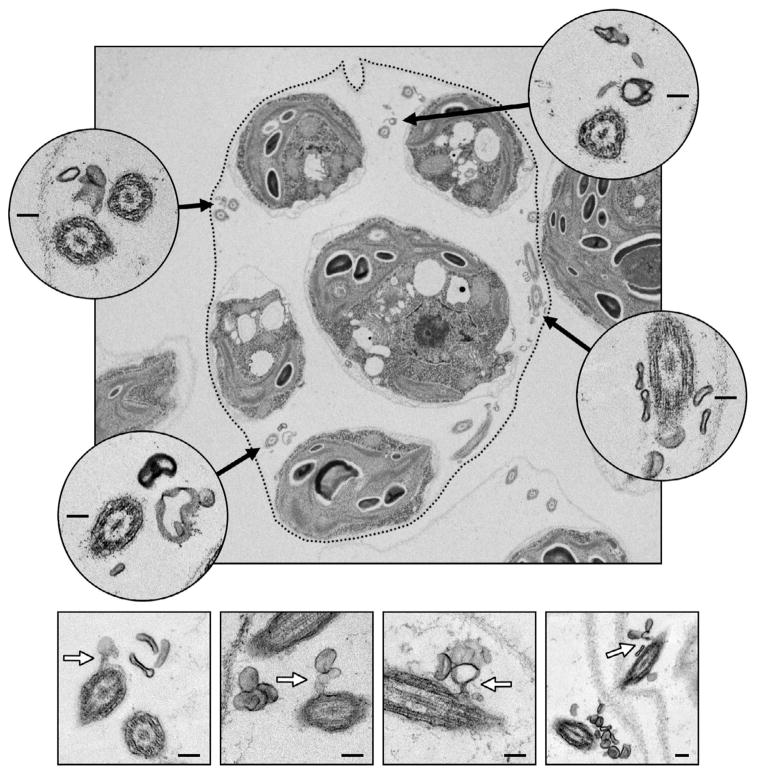
Figure 2. Ciliary ectosomes observed in the mature sporangium by electron microscopy.
In the upper panel, an ultrathin section through a mature sporangium reveals five of the daughter cells within the mother cell wall prior to hatching. The location of the mother cell wall is emphasized by a dotted line. Four insets show higher magnification views of cross sections through flagella (black arrows indicate their position inside the sporangium). Numerous ectosomes are observed clustering around the flagella. The four lower panels display examples of ectosomes caught in the process of budding directly from the membranes of sporangial daughter cell flagella (white arrows). Inset scale bars indicate 100 nm.

