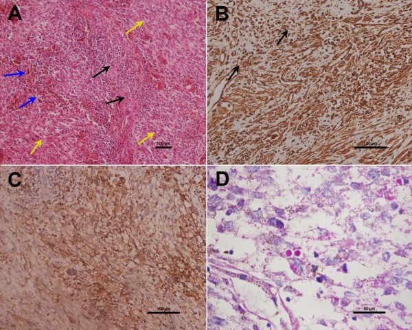
Figure 3.
Histological findings and immunohistochemical expression in UESL. (A) H&E stained sections showing the tumor cells as spindle- or polygonal-shaped with fascicular arrangement (yellow arrows). In addition, multinucleate giant cells (blue arrows), remnants of small bile ducts (black arrows), and chronic inflammatory cells infiltration are shown in the tumor tissue (magnification: 100×). (B, C) Immunohistochemistry analysis of UESL showing positivity for (B) vimentin and (C) AAT (magnification: 200×). In (B), black arrows indicate the coutnerstained cell nuclei. (D) Cells containing eosinophilic cytoplasmic granules staining positive for PAS are shown (magnification: 400×).

