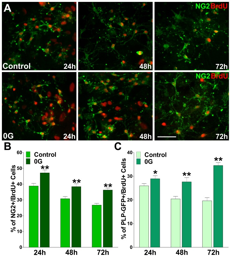
Figure 1. Proliferation of PLP-GFP-expressing OPCs is increased by simulated microgravity.
Mixed glial cultures of PLP-GFP labeled OPCs were treated during 24, 48 and 72 h in 0G. Twenty-four hour pulses of 10 µM bromo-deoxyuridine (BrdU) were begun at 0 h, 24 h and 48 h. After each BrdU pulse, cells were fixed and immunostained with anti-BrdU and anti-NG2 antibodies. (A) Microphotographs showing NG2+/BrdU+ cells at 24, 48 and 72 h. Green: NG2 immunostaining, and Red: BrdU immunostaining. Scale bar = 50 µm. (B and C) The percentage of NG2+/BrdU+ and PLP-GFP+/BrdU+ cells in each experimental condition was compared with respective controls. Values are expressed as mean ± SEM of two independent experiments. *p<0.05, **p<0.01 versus respective control.

