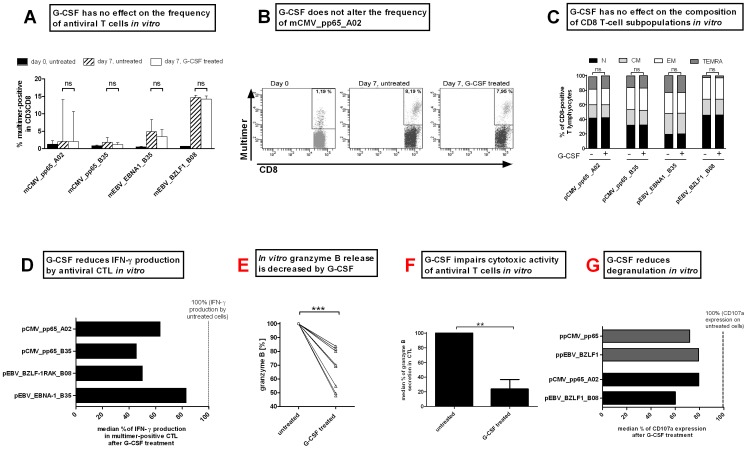
Figure 4. In vitro stimulation with G-CSF does not affect the frequency of antiviral T cells but impairs their functionality.
(A) The frequency of antiviral T cells was measured by HLA-matched multimer staining before (black bars) and 7 days after G-CSF stimulation (untreated: striped bars and G-CSF-treated: white). Medians with range of independent experiments are displayed. (B) Frequency of mCMV_pp65_A02 in a representative CMV-positive donor on day 0 (before stimulation) and after 7 days of pCMV_pp65_A02 stimulation for untreated and G-CSF treated cells. (C) G-CSF treatment did not change the composition of subpopulation within the of CD8+ T-cell population on day 7. (D) IFN-γ secretion as determined by ELISpot analysis after 7 days of peptide stimulation with and without 10 ng/ml G-CSF. The number of IFN-γ secreting units of untreated cells was set to 100%. The number of spot-forming units (sfu) in G-CSF-treated cells is expressed in relation to that in the untreated cells. (E) Granzyme B ELISA, as shown exemplarily for pCMV_pp65_A02 stimulated cells, reveals a reduced secretion of granzyme B after G-CSF treatment. The concentration of secreted granzyme B of untreated cells was set to 100%. (F) G-CSF reduces the cytotoxic activity of antiviral T cells prestimulated over 7 days as shown by granzyme B ELISpot using the pCMV_pp65_A02 loaded T2 cells as target cells. (G) Impaired cytolytic functionality of CD8+ T cells was determined by expression of CD107a on the cell surface. The expression of untreated cells was set to 100%.

