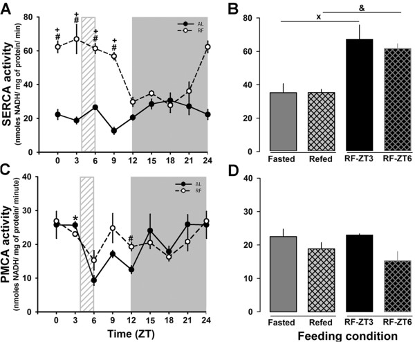
Figure 4.
Hepatic SERCA and PMCA activities Activity of Ca2+-ATPase in microsomal and plasma membrane fractions was measured as described in the methods. The daily profile of SERCA and PMCA activities are shown in panel A and C, respectively. Black circles correspond to AL, and white circles to RF group. The light gray rectangle above x-axis indicates mealtime for the food restricted group (ZT4-ZT6). The corresponding comparisons between feeding conditions (Fasted and Refed) are showed in panel B for SERCA activity and in panel D for PMCA activity. Mean values of 4 independent experiments are shown. Each data point was measured in triplicate. + (p < 0.05) significant difference in the RF group between their time points (1-way ANOVA); # (p < 0.05) significant between AL vs RF (2-way ANOVA). x (p < 0.05) significant difference between Fasted and RF-ZT3 and & (p < 0.05) significant difference between Refed and RF-ZT6 (Student´s t-test).

