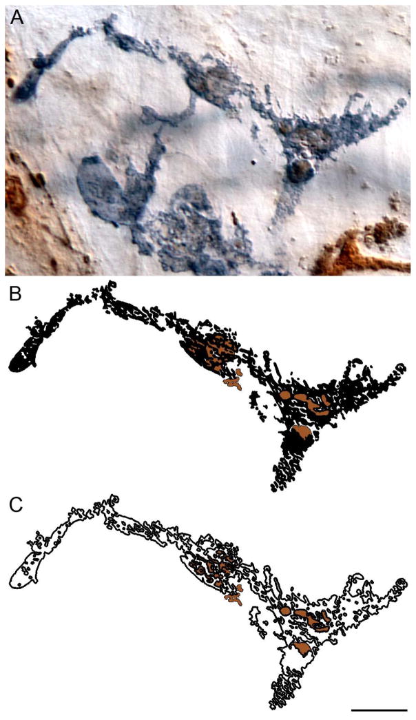
Figure 6.
Macrophages phagocytosed aggregated α-SYNC. A CD163+ macrophage in the duodenum of a 24-month-old rat contained numerous α-SYNC+ aggregates. High power examination (i.e., 100x oil objective) of the macrophage confirmed the presence of the aggregates within the same plane of focus as the macrophage, and therefore the cell’s incorporation of the aggregates, consistent with phagocytosis (A). Some aggregates within the cell were round and well formed, while other aggregates were weakly stained with a flat, diffuse appearance and poorly delineated borders. Panel (B) illustrates in silhouette phagocytosed alpha-synuclein debris (dark brown) within the CD163+ macrophage (black) from panel A; this is further illustrated when only the outline of the macrophage is shown (C). Focus stacking was used to create the extended depth of field image shown in panel A, whereas the illustrations in panels B and C were generated and validated by tracing each plane of focus throughout the z-stack. Scale bar = 10 μm in C (applies to A–C).

