
Figure 2. Multipotent marker expression in Mgp−/− and Mgp−/−;Tie2-Gfptg endothelium.
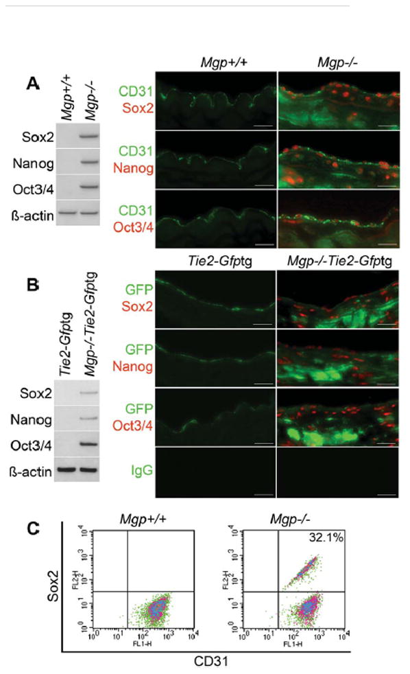
(A) Aortic expression of Sox2, Nanog and Oct3/4 in Mgp+/+ and Mgp−/− mice detected by immunoblotting (left) and immunostaining (right). β-actin was used as control. (B) Aortic expression of Sox2, Nanog and Oct3/4 in Tie2-Gfptg and Mgp−/−;Tie2-Gfptgmice determined by immunoblotting (left) and immunostaining (right). β-actin was used as control. (C) Co-expression of CD31 and Sox2 in enzymatically dispersed CD45-negative aortic cells from Mgp+/+ and Mgp−/− mice, as determined by FACS. Scale bars, 50 μm. Non-specific IgG control showed no staining. Vessel lumen faces upwards in the photos.