
Figure 6. Endothelial origin of multipotent cells in aortas of diabetic Ins2Akita/+ mice.
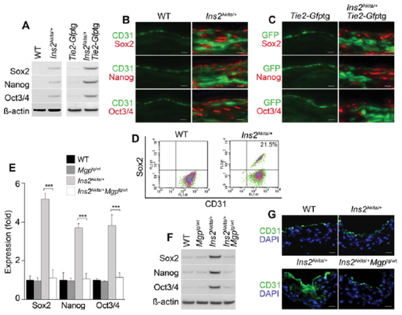
(A) Aortic expression of Sox2, Nanog and Oct3/4 in wild type (WT), Ins2Akita/+, Ins2Akita/+;Tie2-Gfptg and Ins2Akita/+;Tie2-Gfptg mice. β-actin was used as control. (B) Co-expression of CD31 with Sox2, Nanog and Oct3/4 in aortas of Ins2Akita/+ mice detected by immunostaining. (C) Co-expression of GFP with Sox2, Nanog and Oct3/4 in aortas of Tie2-Gfptg and Ins2Akita/+;Tie2-Gfptg mice detected by immunostaining. (D) Co-expression of CD31 and Sox2 in enzymatically dispersed CD45-negative aortic cells from WT and Ins2Akita/+ mice, as determined by FACS. (E–G) Enhanced MGP expression limits aortic expression of Sox2, Nanog and Oct3/4 in Ins2Akita/+ mice, as determined by (E) real-time PCR, (F) immunoblotting (β-actin was used as control), and (G) immunostaining in WT, Mgptg/wt, Ins2Akita/+, and Ins2Akita/+;Mgptg/wt mice. Scale bars, 50 μm. DAPI (blue) was used to visualize nuclei. Vessel lumen faces upwards in the photos.