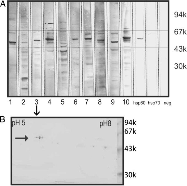
Figure 2.
Western blotting of human retinal proteins with 10 representative sera of autoimmune retinopathy patients. (A) Immunoblotting shows that autoantibodies bind to different retinal proteins within the molecular range of 60 to 70-kDa (boxed). These sera 1–10 were selected to be further analyzed by 2-D western blotting (see Figure 3). (B) A representative 2-D western blot incubated with serum#3 shows 3 molecular forms of 62-kDa protein. Negative control – no primary antibody, positive controls – specific antibodies against human Hsp60 and Hsp70 proteins.

