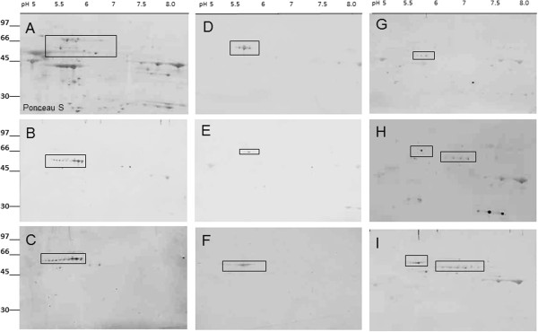
Figure 3.

Representative 2-D western blot analysis of patients sera analyzed by 2-dimensional SDS-PAGE of human retinal proteins; Proteins were first separated by 2-D gel electrophoresis and transferred to a PVDF membrane, then the membrane was stained Ponceau S (A) and destained before incubation with human sera at 1:100 dilution overnight (B-I). Molecular standards are shown on the left and pH range is shown on the top of 2-D gel. The boxed spots on the blots marked the positions of excised proteins from the companion CBB stained gel that was analyzed by mass spectrometry. In B, C, D, and E - sera contained AAbs against hsp60, in F – serum with AAbs against hsp70, and in G, H, I - sera contain mainly AAbs against CRMP-2 and 3. Multiple spot of nearly horizontal spots of the same protein represent isoforms of the same protein.
