
Figure 2.
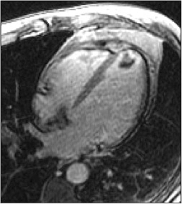
Example of a New Diagnosis. A 32 year-old woman with sickle cell anemia was referred for evaluation of iron overload by T2* imaging, which was normal. However, nearly transmural hyperenhancement (white arrows) was seen in the apical inferior wall on late enhancement imaging, indicative of previously unrecognized myocardial infarction.