Figure 3.
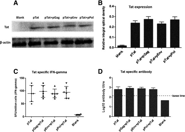
In vitro expression and immune response of Tat. Part A and B showed the Tat expression in 293 T cells. Briefly, 293 T cells (1 × 10⁶ cells per well, and 90% cell viability at least) were transfected with pTat alone, pGag + pTat, pEnv + pTat, pPol + pTat and empty vectors, respectively. Forty-eight hours after transfection, Tat expressions in above-mentioned cell groups were detected by WB method (Figure 3A), and the relative integral optical density of Tat protein in each group was measured (Figure 3B). Empty vector transfection was used as blank control and β-actin was selected as internal control. In part A and B: pTat alone, pGag + pTat, pEnv + pTat, pPol + pTat means that 293 T cells received 4 μg pTat plus 4 μg empty vector, 4 μg pTat plus 4 μg pGag, 4 μg pTat plus 4 μg pEnv, 4 μg pTat plus 4 μg pPol, respectively. Blank: cells were transfected with 8 μg empty vector. Part C and D showed the Tat-specific immune responses. Female BalB/C mice were immunized twice. Two weeks after the final vaccination, mice were sacrificed and the spleens and blood were harvested. Tat-specific T cell and antibody responses were detected with IFN-gamma ELISPOT and IgG ELISA assay, respectively. In part C and D, pTat alone: mice received 50 μg pTat and 50 μg empty vector each time; pGag + pTat/ pEnv + pTat/ pPol + pTat: mice received 50 μg pGag/ pEnv/ pPol and 50 μg pTat each time; Blank: mice received 100 μg empty vector each time.

