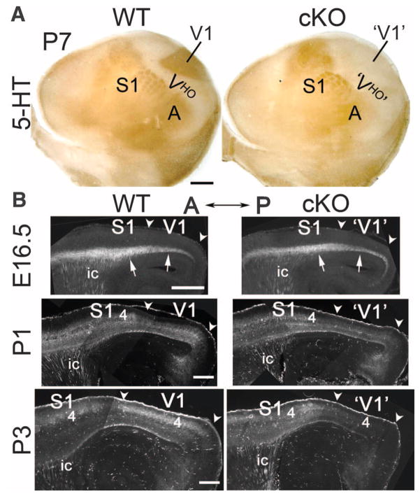
Fig. 1. Selective deletion of geniculocortical TCA projection to V1 in RORα-IRES-Cre floxed COUP-TF1 cKO mice occurs early postnatally.
(A) Geniculocortical projection to V1 is selectively absent in P7 cKO mice. 5-HT immunostaining on tangential sections through layer 4 of P7 WT (COUP-TF1fl/+; RORαCre/+) and cKO (COUP-TF1fl/fl; RORαCre/+) flattened cortices is shown. Rostral is at left and medial at the top. 5-HT staining reveals TCA input from principal sensory thalamic nuclei to primary sensory areas: dLG to V1, VP to S1, and MG to the primary auditory area (A). The 5-HT–negative region surrounding V1 is composed of VHO. In P7 cKO mice, 5-HT immuno-stained geniculocortical TCA input to V1 is absent. 5-HT staining of S1 appeared modestly diminished in cKO as compared to WT mice probably because COUP-TF1 may influence TCA input from VP, which expresses Cre (fig. S1) and exhibits COUP-TF1 deletion (fig. S2), but VP is less affected than dLG by COUP-TF1 deletion (figs. S2 and S4 to S6). (B) Time course of the deletion of geniculocortical TCA projection from dLG to V1 revealed in WT (COUP-TF1fl/+; RORαCre/+; Ai14) and cKO (COUP-TF1fl/fl; RORαCre/+; Ai14) mice crossed to the Ai14 line with the Cre-inducible axon reporter tdTomato. Sagittal sections from E16.5, P1, and P3 WT and cKO cortices, showing TCAs labeled by tdTomato reporter activated by Cre expressed in dLG and VP (anterior to the left, dorsal at the top) are shown. At E16.5, labeled TCAs (arrows) are densely packed in the subplate underlying the cortical plate of nascent V1, with the TCA projection being indistinguishable between WT and cKO mice. At P1, the geniculocortical TCA projection is beginning to invade the overlying CP of V1 in WT mice but is retarded in the cKO mice. By P3, the geniculocortical TCA projection is densely terminating in V1 of WT mice but is virtually absent from V1 in cKO mice. Abbreviations are as follows: 4, layer 4; ic, internal capsule; arrowheads approximate the anterior (A) – posterior (P) extent of nascent V1. Scale bars, 0.5 mm in (A) and 0.2 mm in (B).

